


 18350 Timber Forest Drive, Suite 200
 281-318-7060
 stevie@preciousmemoriesultrasound.com
 Monday 11am to 7:30pm

-
2D Ultrasounds
The Best Way to Determine Your Baby’s Sex Is With 2D Pregnancy Ultrasounds
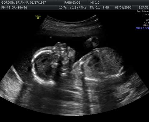
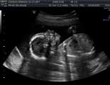
2D Pregnancy Ultrasounds can be done as early as 16 weeks and are the best way to determine your baby’s sex at this stage. 2D ultrasounds are flat black and white moving images of the inside of your body and usually take about 10 minutes. Sound waves travel from the ultrasound transducer through the body and back again to produce a basic image that allows us to view your baby’s internal organs and muscles. This type of ultrasound is useful in detecting things like tumors, polyps or cysts in a major organ. While the images are not true to life, they do produce a clear internal image of your baby which can also help doctors detect and diagnose certain medical issues. 2D ultrasounds are most often used during pregnancy to make sure your baby is healthy and growing well. We also utilize this technology to measure head, stomach, and leg size as well as to monitor the heart rate and movement of the baby.
View Packages
Check Your Baby’s Heartbeat with a 2D Pregnancy Ultrasound
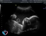
As early as 4 weeks, we can detect and monitor your baby’s heartbeat using the same methods outlined above. In addition to monitoring the health and development of your baby, Pregnancy Ultrasounds use sound waves to create a picture. The traditional ultrasound is a 2D image of a developing fetus and outlines flat-looking images, which can be used to see the baby's internal organs.
2D ultrasounds have been used for decades and have an excellent safety record. Since these devices use non-ionizing radiation, they don't hold the same risks as X-rays, which use ionizing radiation.
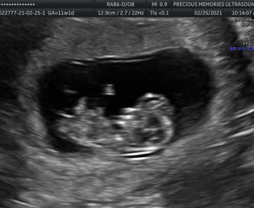
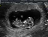
The standard 2D obstetric ultrasound shows a flat, black and white picture on a screen. 2D ultrasounds can be done during any trimester, but are often performed early in gestation to confirm the pregnancy and to help establish the due date. Just four weeks after fertilization, the heartbeat can be picked up on an ultrasound. This scan is usually repeated at 18-20 weeks of gestation to check for normal growth and development, and to reveal the sex of the baby if desired.
Moving the transducer enables numerous planes of viewing, and when the right plane is achieved, as judged by the image on the monitor, a still film can be developed from the recording. To date, Most of the detailed evaluation of fetal anatomy and morphology has been done using 2D ultrasound.
What is a 2D Pregnancy Ultrasound?
A 2D Pregnancy Ultrasound is the standardized procedure used during obstetric ultrasound. 2D ultrasounds are used to produce 2 dimensional images (2D Ultrasounds) in order to show what is happening inside the mother’s body and the baby’s body.
The proper execution of a 2D ultrasound by a trained and experienced professional allows the mother, family members, and healthcare professionals a detailed look at the baby’s gestation, growth, heartbeat, development, and their size can. In addition, the position of the placenta, umbilical cord and lie/position of the baby can also be clearly seen.
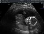
The images from a 2d ultrasound are typically seen in standard black and white and have the same level of detail as a photographic negative. With a 2D ultrasound we will see a variety of images depending on how the baby is lying as well as their position in the uterus.
If you are lucky, they baby might be awake and active during your 2D ultrasound, but keep in mind that it is also likely that your baby will be asleep during this process. Some mothers choose to have a sweet or cold drink before the procedure or “poke” their tummy to stimulate their baby, at Precious Memories, we always advise the Mother to consult her doctor prior to the ultrasound to ensure the well-being of the baby.
Sometimes the images are very clear and it’s easy for parents to interpret the ultrasound images. At other times, this can be a little more difficult. But don’t worry; the sonographer can point out specific organs, features and details of the baby if you’re not sure. They can also freeze frame the images, take photos and label different body parts on the screen. With a little explanation, it will all become clear!